The European virus archive (EVA) is an international non-profit association (AISBL) under the Belgian Law created in March 2025.
EVA is a virology consortium providing the largest collection of viral resources worldwide, with the mission of facilitating virology research and protecting Public Health. By providing academic and private virology researchers with the necessary resources to study viruses and address emerging global threats, EVA addresses critical gaps in pandemic preparedness and response: No central virus collection exists in Europe and there is a need for standardised research using reference material and standardised services for reproducibility in translational research.
Becoming EVA AISBL
Before being an Association, the European Virus Archive benefited from the support of the European Commission through three different grants over the last 15 years:
- EVA (2009-2014), 7th Framework Programme for Research under grant agreement ID 228292
- EVA goes global (2014-2019), Horizon 2020 - Research and innovation under grant agreement ID 653316
- EVA-GLOBAL (2020-2024), Horizon 2020 - Research and innovation under grant agreement ID 871029
EVA AISBL’s Missions
- Gather leading expertise for the collection, amplification, characterisation, standardisation and authentification of human, animal and plant virus resources.
- Develop state-of-the-art technologies for the production of virus-derived products and non-infectious materials for use in diagnostics and research.
- Facilitate access to expertise, resources and services for researchers in academia and industry.
- Support Public Health response and research during viral outbreaks.
EVA AISBL’s Collection
- “One virology concept”: large, diverse and “Fit for Purpose” collection
- Common quality grading and QMS (towards ISO 20387and ISO 13485) and Compliance with Nagoya Protocol
- Easy access to the collection: Online ordering and evaluation, Customer support, ratified MTA, Logistics procedures.
EVA AISBL’s Governance
- General Assembly: Decision-making body
- Executive Board: Managing and executive body
- Director General: Ensures the daily management
- Central Coordinating Unit: Ensures the scientific follow-up, coordinate the activity programs, the daily administration and assists the DG
- Advisory Committees: Provides scientific, ethical, and strategic guidance upon decision of the General Assembly
Documents:
-
Download our Legal Statutes :
 EVA-AISBL statutes
-
Download our Gender Equality Plan:EVA-AISBL Gender Equality Plan
- Download our 2025 Annual Report (Incomming)
Members of the EVA Executive Board
- Hervé BOURHY (Institut Pasteur, France)
- Tatjana AVSIC ZUPANC (Univerza v Ljubljani, Slovenia
- Rémi CHARREL (Aix-Marseille Université, France)
- Boris KLEMPA (Biomedicínske centrum Slovenskej akadémie vied, Slovakia)
- Amber Hartman SCHOLZ (Deutsche Sammlung von Mikroorganismen und Zellkulturen GmbH, Germany)
- Maria Beatrice BONIOTTI (Istituto Zooprofilattico Sperimentale della Lombardia ed Emilia Romagna, Italy)
Director General:
- Bruno COUTARD (Aix-Marseille Université, France)
Governance structure

To contact us:
Access and benefit sharing under the Convention on Biological Diversity and the Nagoya Protocol
The EVA-AISBL community supports fair and equitable benefit sharing as conceived in various United Nations frameworks. The consortium is actively implementing a CBD/Nagoya Protocol compliance strategy.
Privacy policy and GDPR compliance
Personal data is collected when you create an account and when you place an enquiry for any product or service, or when you initiate these processes. Personal data is collected to allow you to personalise your use of the EVA-AISBL website and related services’ (placing enquiries, email notifications, newsletters) and for the delivery of ordered products...
Licence and data reuse
Except where otherwise indicated, original content produced by EVA-AISBL for this website and public EVA Portal catalogue data, including catalogue records and their associated metadata, are licensed under the Creative Commons Attribution 4.0 International (CC BY 4.0) licence. This includes data displayed on web pages or made available through structured formats, feeds, exports and APIs.
This licence permits the licensed content and catalogue data to be copied, extracted, shared, redistributed and adapted for any purpose. Reusers must credit the European Virus Archive – AISBL, link to the licence and the relevant EVA source where reasonably practicable, and indicate whether changes were made. Attribution must not imply endorsement by EVA-AISBL.
Suggested attribution
European Virus Archive – AISBL (EVA), “[title or description of the webpage, catalogue record or dataset]”, available at [source URL], licensed under CC BY 4.0. Where applicable, indicate that changes were made.
Using the EVA logo for attribution
To make the source of reused EVA Portal catalogue data clear and consistent, EVA-AISBL requests that, where appropriate and reasonably practicable, a link to the EVA website and the EVA logo be displayed alongside data obtained from the EVA Portal, including data obtained through structured exports, feeds, APIs or automated extraction.
The following code may be used to display the EVA logo with a link to the EVA website:
<a href="https://www.european-virus-archive.com/">
  <img
    src="https://cdn.european-virus-archive.com/sites/default/files/LOGO_EVA.svg"
    alt="European Virus Archive – AISBL"
    style="max-width: 180px; width: 100%; height: auto;"
  >
</a>
EVA-AISBL authorises use of this logo solely for the purpose of identifying EVA as the source of reused content or catalogue data. The logo must not be modified or used in a way that suggests endorsement, certification, sponsorship or partnership by EVA-AISBL.
The CC BY 4.0 licence does not apply to content identified as belonging to third parties, the EVA name, logos and visual identity, software distributed under another licence, non-public or restricted information, or the physical biological materials and related products described in the catalogue. In particular, images and other media listed in the Image and media credits section below are third-party content and are not covered by EVA-AISBL’s CC BY 4.0 licence, unless the relevant credit explicitly states otherwise.
This general CC BY 4.0 notice does not override a specific licence, copyright notice or rights statement attached to a webpage, file, dataset, image or other content item. Where such a statement is provided, it governs the reuse of that particular item.
Access to and use of physical biological materials and related products remain subject to the applicable access conditions, Material Transfer Agreements, provider requirements, biosafety and biosecurity rules, and Access and Benefit-Sharing obligations. These conditions govern the materials and products themselves and do not change the CC BY 4.0 status of the public EVA Portal catalogue data describing them.
Medias used on this website
Credits
 Electron Microscopy image of the Monkeypox virus
Monkeypox virus directly from a skin lesion sample. Negative contrast. Scale size: 200 nm. Images were taken with a transmission electron microscope JEM-1400 Plus, JEOL, Tokyo, Japan.
 Coronavirus
Produced by the National Institute of Allergy and Infectious Diseases (NIAID), this highly magnified, digitally colorized transmission electron microscopic (TEM) image reveals ultrastructural details exhibited by three, spherical shaped, Middle East respiratory syndrome coronavirus (MERS-CoV) virions.
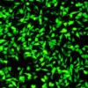 HeLa fluorescence
 Aedes aegypti mosquito
This 2006 image depicted a female Aedes aegypti mosquito as she was obtaining a blood-meal from a human host through her fascicle, which had penetrated the host skin, was reddening in color, reflecting the blood’s coloration through this tubular structure. In this case, what would normally be an unsuspecting host was actually the CDC’s biomedical photographer’s own hand, which he’d offered to the hungry mosquito so that she’d alight, and be photographed while feeding. As it filled with blood, the abdomen became distended, stretching the exterior exoskeletal surface, thereby, causing it to become transparent, allowing the collecting blood to become visible as an enlarging intra-abdominal red mass.
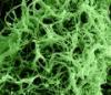 Filamentous Ebola virus particles (SEM)
Produced by the National Institute of Allergy and Infectious Diseases (NIAID), under a magnification of 35,000X, this digitally-colorized scanning electron micrograph (SEM) depicts numerous filamentous Ebola virus particles budding from a chronically-infected VERO E6 cell.